Document Type : Original Article
Authors
1 Graduate Student, School of Veterinary Medicine, Ferdowsi University of Mashhad, Mashhad, Iran
2 Department of Food Hygiene, School of Veterinary Medicine, Ferdowsi University of Mashhad, Mashhad, Iran
3 Department of Clinical Sciences, School of Veterinary Medicine, Ferdowsi University of Mashhad, Mashhad, Iran
4 Department of Pathobiology, School of Veterinary Medicine, Ferdowsi University of Mashhad, Mashhad, Iran
Abstract
Clostridium perfringens is an important cause of bacterial food poisoning worldwide. The disease is caused by C. perfringens enterotoxin (CPE) encoded by cpe gene. The aim of this research was to identify the different types of C. perfringens and the presence of cpe gene in isolated bacteria from broilers’ meat marketed in retail meat shops of Mashhad city in Northeastern of Iran. After isolation of C. perfringens using conventional culture method and confirmation by specific 16S rDNA gene, a multiplex polymerase chain reaction assay with specific primers, were performed for toxin typing of isolates. Clostridium perfringens was isolated from 31 broilers’ meat samples (15.50%) out of 200 samples and for toxin typing the results showed 9 isolates as type A (29.03%) and 22 isolates as type C (70.96%). In this study, cpe-positive C. perfringens were detected in eight isolates of type C (25.00%). Our results indicated that C. perfringens type C is the most common type in broiler chicken carcasses.
Keywords
Main Subjects
Introduction
Clostridium perfringens has been classified into five types (A–E) on the basis of its ability to produce more than one of the major lethal toxins α, β, ε, and ι. Enterotoxin producing C. perfringens (cpe+) type A is reported continuously as one of the most common food poisoning agents worldwide.1
The diarrheic and cramping symptoms of C. perfringens type A food poisoning result from C. perfringens entero-toxin (cpe).2 This toxin is both necessary and sufficient for the enteric virulence of C. perfringens type A food poisoning isolates.2 In vivo production of the enterotoxin is associated with sporulation in the intestine, while in vitro production of enterotoxin is obtained in appropriate culture media.3 Only a small fraction (less than 5.00%) of all C. perfringens isolates, mainly belonging to type A carrying the cpe gene.4 The cpe gene can have either a chromosomal or a plasmid-borne location but is nearly always present on the chromosome of food poisoning isolates.5 There is strong association between type A isolates carrying a chromosomal cpe gene and C. perfringens type A food poisoning is attributable (at least in part) to the exceptional heat resistance of those isolates, which should favor their survival in incompletely cooked or improperly held foods.2 Some type C, D, and E, isolates also carry functional cpe genes on large plasmids.6 Surveys clearly demonstrated that C. perfringens isolates are often present in foods, particularly raw meats and poultry.2,7
For toxin detection, in some laboratories, a serum neutralization test on mice or guinea pigs is employed to determine and diagnose bacterial toxin. This method is tedious, time-consuming, expensive and monovalent. Furthermore, it is improper and unethical to apply it at the expense of laboratory animals.8 According to Timoney et al., enzyme-linked immunosorbent assays (ELISA), proved to be a specific, quick and economical method that may replace the serum neutralization test.9 ELISA utilizes polyclonal antibodies to identify C. perfringens toxins.10 However, its disadvantage is the interaction reaction among the produced antibodies works against the toxins, which may make the identification of toxin types difficult.11 Biochemical tests are also incapable of distinguishing different types of C. perfringens.12 Polymerase chain reaction (PCR) is the most modern practical technology in diagnosing infectious diseases and compared with classical techniques, it is rapid (a few hours) and more reliable.10,13 Various PCR protocols, including multiplex PCR assays, have been established to genotype the C. perfringens isolates with respect to cpa, cpb, etx, iap, cpe and cpb2 genes, encoding the alpha, beta, epsilon, iota, entero and beta 2 toxins, respectively.14-21
As molecular typing of C. perfringens is important for epidemiologic surveys and since there has not been enough information about C. perfringens in broilers’ meat in Iran, the purpose of this study was to determine the incidence and toxin typing of C. perfringens in broilers’ meat collected from retail meat shops in Mashhad city of Iran.
Materials and Methods
Sampling. A total of two hundred samples of broiler carcasses were collected randomly from retail meat shops, using rinse technique for recovering surface bacteria as follows: The broiler carcass was placed in a sterile 1 L plastic bag, 300 mL of phosphate buffer was added. After shaking the bag for 15 sec, the rinse suspension was transferred to laboratory on ice and began bacterial analysis within 1 to 4 hr.
Bacterial isolation. After filtration with sterilized cheese cloth and centrifugation at 4000 rpm for 10 min of each rinsed fluid in two 50 mL falcon tubes, 10 mL of fluid tioglicolate (FTG Difco, Detroit, USA) enrichment medium was added to each pellet. One of those two tubes was heat shocked at 72 ˚C for 20 min before anaerobic incubation at 37 ˚C for 24 hr. Each FTG enrichment culture was streaked onto one plate of nutrient agar containing 10% sheep blood and 40 μg mL-1 neomycin and incubated for 24 hr at 37 ˚C in an anaerobic jar (Merck, Darmstadt, Germany). The plates were examined for typical colonies of C. perfringens. Suspected colonies were subjected to macroscopic examination (shape, size and texture of the colonies on blood agar plates).
Preparation of cell lysates. A single colony of each sample was suspended in 100 μL distilled water, boiled for 10 min and then centrifuged at 10,000 rpm for 10 min. The supernatants were collected carefully and used as template DNA for PCR.
Genus-specific PCR. The identity of the 31 recovered isolates was confirmed as C. perfringens based on the species specific 16S rDNA gene PCR, using specific primers, with oligonucleotide sequence.22
Toxin typing. Six pairs of primers were used to determine the presence of cpa, cpb, iA, etx, cpe16 and cpb2 genes,23 using multiplex PCR technique for all isolates(Table 1). Two strains, C. perfringens CIP 106157 (cpa+, cpe+)and C. perfringens CIP 60.61 (cpa+, cpb+, etx+, cpb2+) obtained from Pasteur Institute Collection (CIP; Paris, France) were used as positive controls. Amplification reactions were carried out in 50 μL volume, containing 5 μL 10x PCR buffer, 5 mM dNTPs, 25 mM MgCl2, 5U of Taq DNA poly-merase, 0.50 mM of each cpa oligo, 0.36 mM of each cpb oligo, 0.36 mM of each cpb2 oligo, 0.52 mM of each iA oligo, 0.44 mM of each etx oligo, 0.34 mM of each cpe oligo, and dH2O. Template DNA (10 μL) was added to the mixture. Amplification was programmed in a thermo-cycler (Model TC3000; Techne, Duxford, UK) as follows: 95 ˚C for 3 min followed by 35 cycles at 94 ˚C for 1 min, 55 ˚C for 1 min, 72 ˚C for 1 min and a final extension at 72 ˚C for 10 min.16 The amplification products were detected by gel electrophoresis in 1.5% agarose gel in 1x TAE buffer, stained with 0.5 μg mL-1 ethidium bromide. Amplified bands were visualized and photographed under UV transillumination.
Results
From total of 200 samples, C. perfringens isolated from 31 samples (15.50%) of broiler chicken carcasses and confirmed by using PCR assay amplifying a specific segment of 16S rDNA gene of C. perfringens.
For toxin typing, the bacterial isolates were analyzed by multiplex PCR assay using specific primers in order to determine the presence of cpa, cpb, iA, etx, cpe and cpb2 genes. PCR results corresponding to positive and negative controls are displayed in Figure 2. Out of 31 C. perfringens isolates, 9 (29.03%) isolates were determined as type A (carrying the alpha toxin gene). From these 9 isolates, 4 (44.40%) were determined as simple type A (carrying neither the cpe nor cpb2 gene) and 5 (55.50%) isolates were determined as hetero-geneous types (carrying cpb2 gene) but none of the isolates were found to carry both the cpb2 and cpe genes. As the dominant type, 22 isolates (70.96%) were determined as type C (Fig. 1).
The most important result from multiplex PCR analysis was detection of the cpe gene in eight isolates of C. perfringens which all of them belonged to type C (Fig. 2).
Table 1. Primers for 16S rDNAgene, cpa, cpb, etx, iA, cpe and cpb2 toxin genes detection.
|
Target gene |
Primer sequences (5`-3`) |
Product length (bp) |
References |
Annealing temperatures |
|
16S rDNA |
AAAGATGGCATCATCATTCAAC TACCGTCATTATCTTCCCCAAA |
279 |
22 |
53 ˚C |
|
cpa |
GCTAATGTTACTGCCGTTGA CCTCTGATACATCGTGTAAG |
324 |
16 |
55 ˚C |
|
cpb |
GCGAATATGCTGAATCATCTA GCAGGAACATTAGTATATCTTC |
196 |
16 |
55 ˚C |
|
etx |
GCGGTGATATCCATCTATTC CCACTTACTTGTCCTACTAAC |
655 |
16 |
55 ˚C |
|
iA |
ACTACTCTCAGACAAGACAG CTTTCCTTCTATTACTATACG |
446 |
16 |
55 ˚C |
|
cpe |
GGAGATGGTTGGATATTAGG GGACCAGCAGTTGTAGATA |
233 |
16 |
55 ˚C |
|
cpb2 |
AGATTTTAAATATGATCCTAACC CAATACCCTTCACCAAATACTC |
567 |
23 |
55 ˚C |
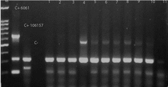
Fig.1. Agarose gel electrophoresis of PCR products obtained from C. perfringens isolated from broiler carcass samples. Lane M: Marker (DNA ladder, 100 bp); C+ CIP 60.61(cpa+, cpe+) and C+106157 (cpa+, cpb+, etx+, cpb2+): Positive controls; C-: Negative control; Lane 4: C. perfringens type A isolate; Lanes 1, 2, 3, 5, 6, 7, 8, 9, 10: C. perfringens type C isolates.
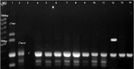
Fig. 2. PCR identification of cpegene. Lane M: Marker (DNA ladder, 100 bp); Lane 1: C+ CIP 60.61 C. perfringens(cpa+, cpb+, etx+, cpb2+) and lane 2: C+ CIP 106157: Positive controls;Lane 3 negative control; Lanes 4,5,6,7,8,9,10,11: C. perfringens type C isolates with cpe gene; Lanes 12 , 13 and 14: C. perfringens type A isolates.
Discussion
Food poisoning caused by C. perfringens is among the common illnesses resulting from the consumption of contaminated food and it has been firmly established that enterotoxin produced in the intestine following sporulation of ingested vegetative cells is responsible for the illness caused by C. perfringens.24
In Norway in the 1990s, C. perfringens was registered as the most common cause of food poisoning.13 The prevalence in other countries, such as Japan, the US and the UK, is also high.13 In England and Wales, C. perfringens was the second most frequently reported organism associated with food borne outbreaks of intestinal disease in the 1990s.25,26 The vehicles of infection are typically meat and poultry products.2 A survey by Lin and Labbe demonstrated these foods to be the most heavily contaminated with C. perfringens isolates.27
According to a report published in 2013, per capita consumption of poultry in Iran is approximately 25.16 kg (higher than the world average consumption).28 Pilgrimage and tourist attractions of the Mashhad city in Northeastern of Iran provides it with nearly 32 million pilgrims every year,29 and the amount of food which serves for this population, reveals the importance of foodborne illness in this city.
In recent decades, many surveys have been conducted on the incidence of C. perfringens in raw and processed meat and poultry.7 Sperner et al. reported that 20.00% of the fish and broiler meat samples as positive for C. perfringens.30Higher incidence of C. perfringens has been reported by Miwa et al. in raw chickens meat as 84.00% and by Nowell et al. as 66.00%.31,32 Wen and McClane also reported the incidence of C. perfringens in 38.00% of raw chicken meats.2 In the present study, the total incidence of C. perfringens in broiler carcasses was 15.50%. The different results may be due to different meat processing methods or the method of sampling and bacterial isolation. In our study, the sampling method was rinsing the whole carcass, and the bacterial isolation method was enrichment in FTG medium followed by selective plating on sheep blood agar, and finally DNA extraction from suspected colonies and confirmation by PCR method.
Detection of C. perfringens toxin types and subtypes is critical for a better understanding of the epidemiology of C. perfringens infections and may be helpful in the development of effective preventive measures. The typing of C. perfringens strains was originally established based on neutralization of the pathological effect of each major toxin, both trypsin treated and untreated, with appropriate antisera in laboratory animal models.33 In diagnostic laboratories, this differentiation has been replaced by enzyme-linked immunosorbent assays (ELISA).34 Although ELISA allows reliable typing of C. perfringens isolates, the options for subtyping are limited. For example, so far no ELISA is available to detect the β2-toxin. In addition, high levels of enterotoxin have been shown only to be present during sporulation.35 As a consequence, sporulation of C. perfringens isolates has to be induced via specific cultivation methods to detect enterotoxin producing strains.11 These problems have been solved by genotyping of C. perfringens isolates. Various PCR protocols including multiplex PCR assays have been established to genotype C. perfringens isolates. Sensitivity and specificity are the two main characteristics of an efficient and practical technique existing in PCR. Rapidity is one of the major advantages of this method, so that bacterium identification and type determination lasts no longer than four hours. Hence, the toxicogenic strain in the sample can be identified by means of a rapid evaluation by PCR before it produces toxin. Genotyping of C. perfringens by PCR is a rapid and useful method and the use of a PCR variant, multiplex PCR, has enabled the simultaneous detection of the main toxins with the consequent saving of time.
Several studies reported that type A is the predominant type in poultry. The enterotoxins of type A have been reported to cause food-borne illness in humans.2,36 In contrary, our results showed that type C of C. perfringens was the most prevalent type in broiler meat samples. The same results has been reported by Poursoltani et al. which detected all 180 isolates of C. perfringens from wing, neck, liver and gizzard of broiler chickens in Mashhad, as type C by multiplex PCR.37
Only a small fraction (1.00 to 5.00%) of all C. perfringens isolates, mainly belonging to type A, carry the cpe gene.38 Intact cpe genes can also be found in some type C, D and E strains.6 Results of the present study showed that enterotoxigenic strains of C. perfringens were 25.00% which all of them belonged to type C isolates. Miwa et al. found that enterotoxigenic strains of C. perfringens were present in an average of 12.00% of poultry samples.31 Singh et al. reported the incidences of enterotoxigenic C. perfringens in 15.50% of poultry meat.39
Type C strains of C. perfringens are the only non type A strain that cause human disease,40 which is referred to as enteritis necroticans, also known as pigbel.41 Food poisoning by type C of C. perfringens is lethal in 25.00% of cases.25 The symptoms of C. perfringens type C food poisoning (necrotic enteritis) in human, start with abdominal pain and bloody diarrhea, and are followed rapidly by necrosis of the small intestine, caused mainly by the beta toxin, with contributions of additional toxins (cpe has been proposed as a possible contributor to the pathogenesis of human pigbel).25
In this study, all of the strains were identified as type A and C. The absence of types B, D and E is probably due to the origin of the samples which were broiler meats. In a study with samples of different origins, Songer and Meer reported 92.70% of isolate as type A; 0.10% of as type B; 4.50% of as type C; 2.10% as type D and 0.60% as type E.15 According to our results, it can be concluded that type C is the most predominant type in this region, and because this type causes more deadly illness in human, further investigations are required with larger sample sizes and more geographic distribution in Iran.
Acknowledgements
This research was funded by a grant (No. 28440) from the Research Council of the Ferdowsi University of Mashhad. We would like to thank Mr. Ali Kargar and Mrs. Samira Khajenasir for their assistance in laboratory works.
- Adak GK, Long SM, O’Brien SJ. Trends in indigenous foodborne disease and deaths, England and Wales: 1992 to 2000. Gut 2002; 51: 832-841.
- Wen Q, Mc Clane BA. Detection of enterotoxicogenic Clostridiumperfringens Type A isolates in American retail foods. Appl Environ Microbiol 2004;70(5): 2685-2691.
- Aguileraa MO, Stagnittab PV, Micalizzi B, et al. Prevalence and characterization of Clostridium perfringens from spices in Argentina. Anaerobe 2005; 11: 327-334.
- Smedley JG, Fisher DJ, Sayeed S, et al. The enteric toxins of Clostridium perfringens. Rev Physiol Biochem Pharmacol 2004; 152: 183-204.
- Lindström M, Heikinheimo A, Lahti P, et al. Novel insights into the epidemiology of Clostridiumperfringens type A food poisoning. Food Microbiol 2011; 28: 192-198.
- Miyamoto K, Li J, Mc Clane BA. Enterotoxigenic Clostridium perfringens: Detection and Identification. Microbes Environ 2012: 27: 343-349.
- Labbe RG, Lund BM, Baird Parker TC, et al. Clostridium perfringens. In: The microbiological safety and quality of food. Vol II. Gaithersburg, USA: Aspen. Publishers, Inc., 2000; 1110-1135.
- Piatti M, Ikuno AA, Baldassi L. Detection of bovine Clostridium perfringens by polymerase chain reaction. J Venom Anim Toxins 2004; 10: 154-160.
- Timoney JF, Gillespie JH, Scott FW, et al. Hagan and Bruner’s microbiology and infectious diseases of domestic animals. 8th ed. Ithaca, USA: Comstock Publishing Associates 1988; 214-240.
- Baron EJ, Finegold SM, Martin WJ. Organisms encountered in the urinary tract. In: Bailey and Scott’s diagnostic microbiology. St. Louis, USA: Mosby 1990; 49-194.
- Baums CG, Schotte U, Amtsberg G, et al. Diagnostic multiplex PCR for toxin genotyping of Clostridium perfringens isolates. Vet Microbiol 2004; 100: 11-16.
- Ahsani MR, Mohammadabadi MR, Shamsaddini MB. Clostridium perfringens isolate typing by multiplex PCR. J Venom Anim Toxins 2010; 16: 573-578.
- Brynestad S, Granum PE. Clostridium perfringens and food borne infections. Int J Food Microbiol 2002;74: 195-202.
- Daube G, Simon P, Limbourg B, et al. Hybridization of 2,659 Clostridium perfringens isolates with geneprobes for seven toxins and for sialidase. AmJ Vet Res 1996; 57: 496-501.
- Songer JG, Meer RR. Genotyping of Clostridium perfringens by polymerase chain reaction is a useful adjunct to diagnosis of clostridial enteric disease in animals. Anaerobe1996; 2: 197-203.
- Meer R, Songer JG. Multiplex polymerase chain reaction assay for genotyping Clostridium perfringens. Am J Vet Res 1997; 58: 702-705.
- Yoo HS, Lee SU, Park KY, et al. Molecular typing and epidemiological survey of prevalence of Clostridium perfringens types by multiplex PCR. J Clin Microbiol 1997;35: 228-232.
- Kadra B, Gouillou JP, Popoff M, et al. Typing of sheep clinical isolates and identification of enterotoxigenic Clostridium perfringens strains by classical methods and by polymerase chain reaction (PCR). FEMSImmunol Med Microbiol 1999; 24: 259-266.
- Kanakaraj R, Harris DL, Songer JG, et al. Multiplex PCR assay for detection of Clostridium perfringens in feces and intestinal contents of pigs and in swine feed. Vet Microbiol 1998; 63: 29-38.
- Augustynowicz E, Gzyl A, Slusarczyk J. Molecular epidemiology survey of toxinogenic Clostridium perfringens strain types by multiplex PCR. Scand J Infect Dis 2000; 32: 637-641.
- Garmory HS, Chanter N, French NP, et al. Occurrence of Clostridium perfringens b2-toxin amongst animals, determined using genotyping and subtyping PCR assays. Epidemiol Infect 2000; 124: 61-67.
- Wu J, Zhang W, Xie B, et al. Detection and toxin typing of Clostridium perfringens in formalin-fixed, paraffin-embedded tissue samples by PCR. J Clin Microbiol 2009; 47: 807-810.
- Bueschel DM, Jost BH, Billington SJ, et al. Prevalence of cpb2, encoding beta2 toxin, in Clostridium perfringens field isolates: Correlation of genotype with phenotype. Vet Microbiol 2003; 94: 121-129.
- Duncan C, Strong DH. Improved medium for sporulation of C. perfringens. Applied Microbiol 1968; 16: 82-89.
- Van Immerseel F, De Buck J, Pasmans F, et al. Clostridium perfringens in poultry: An emerging threat for animal and public health. Avian Pathol 2004; 33:537-549.
- Kessel AS, Gillespie IA, O’Brien SJ, et al. General outbreaks of infectious intestinal diseases linked with poultry, England and Wales, 1992-1999. Commun Dis Public Health 2001; 4: 171-177.
- Lin YT, R Labbe.Enterotoxigenicity and genetic relatedness of Clostridium perfringens isolates from retail foods in the United States. Appl Environ Microbiol 2003; 69: 1642-1646.
- Livestock and poultry. Ministry of Agriculture. Center for Information and Communication Technology. Agricultural statistics, second volume. Available at http://ibs.agri-jahad.org. Accessed Oct 01, 2014.
- Ghasemi M. Analysis of the development to religious tourism in the city of Mashhad. 1st international conference on religious tourism and pilgrimage culture, Islamic Republic of Iran. 2011; 6-10.
- Sperner B, Schalch B, Eisgruber H, et al. Molecular methods for the analysis of Clostridium perfringens relevant to food hygiene. FEMS Immunol Med Microbiol 1999; 24: 281-286.
- Miwa N, Nishina T, Kubo S, et al. Amount of entero-toxigenic Clostridium perfringens in meat detected by nest PCR. Int J Food Microbiol 1998; 42: 195-200.
- Nowell N, Poppe C, Parreira VR, et al. Clostridium perfringens in retail chicken. Anaerobe 2010; 16: 314-315.
- Sterne M, Batty I. Pathogenic clostridia. London, UK: Butterworths 1975: 79-122.
- Nagahama M, Kobayashi K, Ochi S, et al. Enzyme-linked immunosorbent assay for rapid detection of toxins from Clostridium perfringens. FEMS Microbiol Lett 1991;68: 41-44.
- Czeczulin JR, Hanna PC, Mc Clane BA. Cloning, nucleotide sequencing, and expression of the Clostridium perfringens enterotoxin gene in Escherichia coli. Infect Immun 1993; 61: 3429-3439.
- Engstrom BE, Fermer C, Lindberg A, et al. Molecular typing of isolates of Clostridiumperfringens from healthy and diseased poultry. Vet Microbiol 2003; 94: 225-235.
- Poursoltani M, Razmyar J, Mohsenzadeh M, et al. Toxinotyping of Clostridium perfringens strains isolated from packed chicken portions. Iran J Med Microbiol 2014; 8(1):9-17.
- Kokai-Kun JF, Songer JG, Czeczulin JR, et al. Comparison of Western immunoblots and gene detection assays for identification of potentially enterotoxigenic isolates of Clostridium perfringens. J Clin Microbiol1994; 32: 2533-2539.
- Singh RV, Blilegaonkar KN, Agarwal RK. Studies on occurrence and characterization of Clostridium perfringens from select meats. J Food Safety 2005; 25: 146-156.
- Mc Clane BA, Uzal FA, Miyakawa MF, et al. The enterotoxic clostridia. In: Dworkin M, Falkow S, Rosenburg E, et al. (Eds). The prokaryotes. 3rd ed. New York, USA: Springer 2006; 688-752.
- Johnson S, Gerding DN. Enterotoxemic infections. In: Rood JI, Mc Clane BA, Songer JG, et al. (Eds). The clostridia: Molecular biology and pathogenesis. London, UK: Academic Press 1997; 117-140.
)