Document Type : Clinical Report
Authors
1 Department of Pathobiology, Faculty of Veterinary Medicine, Lorestan University, Khorram Abad, Iran
2 Department of Pathobiology, School of Veterinary Medicine, Shiraz University, Shiraz, Iran
Abstract
Leydig cell tumor as a sex-cord stromal tumor is a relatively uncommon ovarian tumorin bitch. A 10-year-old female German Shepherd dog was presented because of protrusion of a large tumor-like mass of 16 × 14 × 7 cm in dimensions from her vagina. After stabilization of the patient, the mass was removed surgically and concurrent ovariohysterectomy was also performed. Macroscopically, the healthy tissue of the right ovary was totally replaced by a homogeneous, brown and firm mass. The neoplasm was well-circumscribed and nodular and it was clearly demarcated from the healthy tissue. Histological examination revealed the presence of solid sheets and acinar structures composed of polyhedral to elongated cells. The neoplastic cells had large, eosinophilic, and vacuolated cytoplasms with round to oval nuclei and expressed vimentin on immunohistochemical examination. These gross, microscopic and immuno-histochemical features are characteristics of ovarian Leydig cell tumor.
Keywords
Main Subjects
Introduction
Ovarian tumors are relatively uncommon in bitch. Their frequency ranges between 0.5 and 1.2% with an average of 1.0%, but their incidence is probably under-estimated.1 According to the world health organization, the ovarian tumors can histologically be classified as granulose theca cell stromal tumor, Sertoli-stromal cell tumor, sex cord tumor with annular tubules, gynandro-blastoma, unclassified and steroid cell tumors. The Sertoli-stromal cell tumor consists of Sertoli-Leydig cell tumors, as well as pure Sertoli cell or pure Leydig cell tumors. Leydig cell tumors account for 15.0% to 20.0% of the steroid cell tumors in human.2,3 The Leydig cell tumors of the ovary are composed of cells resembling those of the corpus luteum in all species. These tumors consist of multiple lobules of neoplastic cells separated by a well-vascularized connective tissue stroma. The neoplastic cells are polygonal with abundant granular eosinophilic cytoplasm containing lipid vacuoles.4 The present report describes the histopathological and immunohistochemical features of a Leydig cell tumor in a German Shepherd bitch.
Case Description
A 10-year-old female German Shepherd dog was admitted due to protrusion of a large mass of 16 × 14 × 7 cm in dimensions (Fig. 1) from her vagina which resulted in severe depression, lethargy and anorexia. During clinical examinations, pale mucous membrane, tachycardia, tachypnea and distention of the bladder, probably due to compression of the urethra by the mass were detected. After stabilization of the patient the mass was removed surgically and concurrent ovariohysterectomy was also performed. On gross examination, the healthy tissue of the right ovary was totally replaced by a tumor like mass. The mass was brown, homogeneous, firm and elastic at palpation. It was well-circumscribed, nodular and clearly demarcated from the healthy ovary.

Fig. 1. Leydig cell tumor in a 10-year-old bitch. A large mass measuring 16 × 14 × 7 cm protruded from the vagina.
The appropriate tissues were fixed in 10% neutral buffered formalin, dehydrated in graded ethanol, cleared in xylene and embedded in paraffin wax. Sections of 5 µm thicknesses were stained with hematoxylin and eosin (H & E) and studied by a compound light microscope. Serial sections were subjected to immunohistochemistry with primary monoclonal antibodies specific for pancyto-keratin, epithelial membrane antigen (EMA), S-100, inhibin, calretinin, synaptophysin, neuron-specific enolase (NSE), chromogranin and vimentin. All primary anti-bodies were from Novocastra Laboratories (Newcastle, UK). Labelling was detected with an avidin-biotin conjugate (ABC) procedure.
Histological examination revealed presence of the solid sheets and acinar structures composed of polyhedral to elongated cells. Broad bands of fibrovascular stroma divided the encapsulated neoplastic mass into lobules of different sizes and shapes. The neoplastic cells had large, eosinophilic, and vacuolated cytoplasms and round to oval nuclei containing small nucleoli (Fig. 2). The single or multiple clear vacuoles were of different sizes and optically empty (Fig. 3).

Fig. 2. Microscopic examination reveals presence of solid sheets and acinar structures of Leydig cell tumor with polyhedral to elongated, vacuolated cells (H & E, Bar: 125 µm).
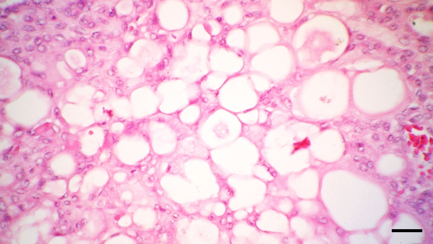
Fig. 3. The neoplastic cells have eosinophilic and round to oval nuclei with single or multiple cytoplasmic vacuoles (H & E, Bar: 30 µm).
In some parts of the tumor, angiogenesis was prominent which at some instances resulted in small hemorrhagic foci. The neoplastic cells labelled cytoplasmic positivity for vimentin (Fig. 4), but did not express any other marker. These immunohistochemical findings allowed confirming the diagnosis of Leydig cells tumor.

Fig. 4. The tumor cells show cytoplasmic positivity for vimentin (IHC, Bar: 30 µm).
Discussion
Canine ovarian neoplasms are uncommon.5,6 They occur more frequently in older, multiparous bitches or in bitches with ovarian remnant syndrome.7 Interstitial-cell tumors are relatively more common in male testes but the ovarian interstitial-cell tumors are rare in all species.8 Unlike the male dogs, the tumor is not common in ovaries of females. Leydig stromal tumor is usually benign, unilateral and metastasis of the tumor is very uncommon.9,10 During examination of the present case, pale mucous membrane, tachycardia, tachypnea and distension of the bladder probably due to pressure of the mass on urethra were detected. No details of the clinical signs have been provided in the ovarian interstitial-cell tumors that have previously been reported.11,12
An interstitial-cell tumor of the ovary resembles a corpus luteum both grossly and histologically.13 Micro-scopically, the cells typically have abundant eosinophilic cytoplasm; however, some cells may have vacuolated cytoplasm indicative of lipid. The nuclei are typically round with a single small nucleolus. The histologic description of the tumors in this case was similar to those described in cattle, eland, alpaca and dog.13-15
Most Leydig cell tumors show positive staining for vimentin.15-18 Other staining reactions that have been reported include staining for chromogranin, synapto-physin, S100 and cytokeratin.16 In the present report, the neoplastic cells labelled cytoplasmic positivity for vimentin. Ovariohysterectomy is a prudent method for preventing many diseases in dogs and cats, such as pyometra, mammary and ovarian tumors. In the present case, after stabilization of the patient, the mass was removed surgically and concurrent ovariohysterectomy was also performed.
Leydig cell tumor is a very rare neoplasma in the bitch. Veterinary practitioners should enlighten pet owners regarding the possibility of this neoplasm.19
Acknowledgements
The authors would like to thank Mr. Ghasem Yousefi (Department of Pathobiology, School of Veterinary Medicine, Shiraz University, Shiraz, Iran) for his technical assistance.
- Dow C. Ovarian abnormalities in the bitch. J Comp Pathol 1960; 70: 59-69.
- Scully RE. International histological classification of tumors. Histological typing of ovarian tumors. Berlin, Germany: Springer 1999; 24-25.
- Gershenson DM, Young RH. Ovarian sex cord-stromal tumors. In: Hoskins WJ (Ed). Principles and practice of gynecologic oncology. 4th ed. Philadelphia, USA: Lippincott Williams & Wilkins; 2005:1024.
- Mc Entee MC. Reproductive oncology: Clinical techniques. J Small Anim Pract, 2002; 17, 133-149.
- Arnold S. Ovarian tumors. In: Practical course of the dog clinic [German]. Niemand HG, Suter PF (Eds). 9th ed. Berlin, Germany: Parey 2001; 926-927.
- Ortega-Pacheco A. Reproduction of dogs in the tropics with special reference to the population structures, reproductive patterns and pathologies and a non-surgical castration alternative. Doctoral thesis. Swedish University of Agricultural Sciences, Uppsala 2006.
- Matsuu A, Hashizume T, Kanda T, et al. A case of persistent Müllerian duct syndrome with Sertoli cell tumour and hydrometra in a dog. J Vet Med Sci 2009; 71(3): 379-381.
- Takeuchi S, Ishihara N, Ohbayashi C, et al. Stromal Leydig cell tumor of the ovary: Case report and literature review. Int J Gynecol Pathol 1999; 18:178-182.
- Roth LM. Sex cord stromal tumor of the ovary. In: Fox H, Wells M (Eds). Haines and Taylor, obstetrical and gynecological pathology. 5th ed. Edinburgh, UK: Churchill Livingstone 2003; 745-765.
- Colombo Ni, Parma Ga. Ovarian sex cord stromal tumors. In: Gershenson DM, Mcguire Mc, Gore M, et al (Eds). Gynecologic cancer. 2nd ed. USA: Elsevier 2005; 503-507.
- Gelberg HB, McEntee K. Feline ovarian neoplasms. Vet Pathol 1985; 22: 572-576.
- Gilbert R, Kutzler M, Valentine A, et al. Hyper-androgenism from an ovarian interstitial-cell tumor in an alpaca. J Vet Diagn Invest 2006; 18: 605-607.
- Owston MA, Ramos-Vara JA. Histologic and immuno-histochemical characterization of a testicular mixed germ cell sex cord-stromal tumor and a Leydig cell tumor in a dog. Vet Pathol 2007; 44: 936-943.
- Nyska A, Waner T, Goldstein J, et al. interstitial (Leydig) cell tumor in eland (Taurotragus oryx). J Wildl Dis 1994; 30(2): 291-294.
- Busam KJ, Iversen K, Coplan KA, et al. Immuno-reactivity for A103, an antibody to melan-A (Mart-1), in adrenocortical and other steroid tumors. Am J Surg Pathol 1998; 22(1): 57-63.
- Iczkowski KA, Bostwick DG, Roche PC, et al. Inhibin A is a sensitive and specific marker for testicular sex cord- stromal tumors. Mod Path 1998; 11(8): 774-779.
- McCluggage WG, Shanks JH, Whiteside C, et al. Immunohistochemical study of testicular sex cord stromal tumors, including staining with anti-inhibin antibody. Am J Surg Pathol 1998; 22(5): 615-619.
- Augusto D, Leteurtre E, De La Taille A, et al. Calretinin: A valuable marker of normal and neoplastic Leydig cells of the testis. Appl Immunohistochem Mol Morphol 2002; 10(2): 159-162.
- Vissiennon TH, Schmidt TH, Schneider E, et al. Sertoli Leydig cell tumor in a bitch, 10 years after spaying. Tierarztl Prax Kleintiere 2010; 38: 399-402.
)