Vet Res Forum
Document Type : Original Article
Authors
1 Department of Pathobiology, Faculty of Veterinary Medicine, Urmia University, Urmia, Iran
2 Department of Food Hygiene and Quality Control, Faculty of Veterinary Medicine, Urmia University, Urmia, Iran
Keywords
Introduction
Cryptosporidium an obligate intracellular protozoan parasite is a frequent cause of intestinal, gastric or respiratory cryptosporidiosis in a wide range of animals and humans hosts worldwide.1 Some of the zoonotic Cryptosporidium species usually causes self-limiting diarrhea in humans and animals. Cryptosporidiosis was reported for the first in 1971,2 as well as from Iranian cattle in 1984,3 and increasingly in a range of hosts.1,4-7
Over the past two decades, cattle have been identified as a common reservoir host for Cryptosporidium species. Currently, 20 different species of Cryptosporidium has been reported which C. parvum, C. bovis, C. ryanae, Cryptosporidium deer-like genotype, and C. andersoni are considered as cattle adapted.8 Of those, the intestinal specie C. parvum has zoonotic potential and is a frequent cause of human cryptosporidiosis.9,10 In terms of economic losses, C. parvum is considered as the most important species among cryptosporidial agents.11 Cryptosporidium andersoni as an abomasal parasite has been associated with reduced milk yield in dairy cattle and decreased weight gain in post weaned calves.12
According to Xiao et al., a high diversity of Crypto-sporidium genus based on several molecular markers, the 18S rRNA gene, has been shown by multilocus DNA analysis.10 In Iran, molecular detection of Cryptosporidium in humans has been undertaken by several researchers.11,13,14 While, to date, the molecular study for confirming cattle Cryptosporidium infection has not been investigated in this part of Iran. Additionally, the nationwide epidemiological survey is essential for the exact knowledge of infection status of C. parvum and C. andersoni in the country. Therefore, it is important to determine bovine Crypto-sporidium infection and major risk factors in order to screen C. parvum and C. andersoni harboring in cattle herds of northwestern Iran.
Materials and Methods
Study area. The study area (West Azarbaijan province, WAP) is located in northwest of Iran with two rainy seasons, the first from March to May and the second in October-November. The study was carried out in the mountainous, mountainside and plain areas of Urmia suburb covering 355 villages that fall within a radius of 20 to 125 km of the city center (Fig. 1). According to the Iranian Veterinary Organization, an average population of eight million cattle is distributed in Iran; the WAP has approximately 6.1% of these cattle.15
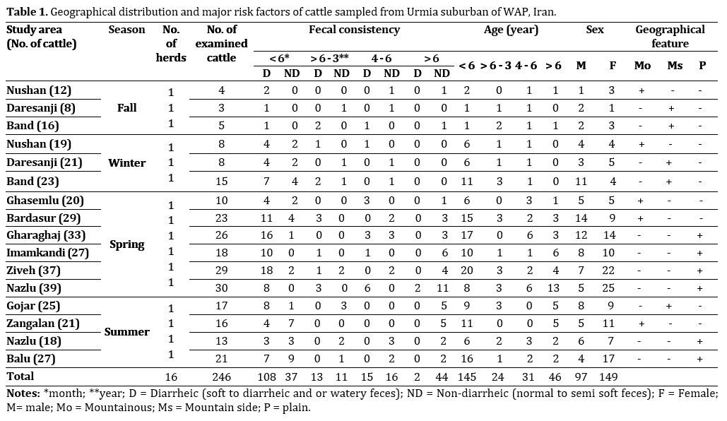
Animals. During the course of the study (September 2010 to August 2011), a total number of 101 cattle and 145 calves from 16 herds (375 cattle) were randomly selected from cattle herds in Urmia suburban of WAP. The herds examined were raised following traditional husbandry practices, with animals being mainly cross-breeds and indigenous crossbred. The major risk factors for Cryptosporidium infection were host, Cryptosporidium species, and environmental factors (Table 1). The cattle were divided into four age groups, numbered and subjected to clinical examination. The age was estimated on the basis eruption of permanent incisor teeth.16 The consistency (Fc) of fecal specimens (D or ND) were recorded. The sample size for determining prevalence was estimated based on the formula (expected prevalence 30%, level of confidence 95%, and precision 5%) presented by Thrusfield.16

Fig. 1. Map of West Azarbaijan province (WAP) showing the places with samples of cattle herd examined for infection.
Sample processing and examination. In each farm and herd, an amount of 25 g fresh fecal samples was collected directly from the rectum of each individual cattle and or calves and fecal smears were prepared. The Cryptosporidium oocysts were initially screened using the modified Ziehl-Neelsen staining method.18 The parasite species was identified by morphologic and morphometeric criteria which measured at 1000× magnification. Morphologically, C. parvum (5.0 × 4.5 µm) oocysts were discriminated from C. andersoni oocysts (7.6 × 5.6 µm).19
Molecular procedures. Since, the modified Ziehl-Neelsen staining is a non-specific staining procedure the PCR procedure was performed to specify genus Cryptosporidium. For this purpose, oocysts were purified from fecal specimens of infected cattle using sucrose gradients as described by Arrowood and Sterling 20 and subjected to molecular analysis. To rupture Crypto-sporidium oocysts, 10 times freeze-thaw cycles were performed using liquid nitrogen.21 Genomic DNA was extracted by modified phenol-chloroform method using cetyltrimethylammonium bromide (CTAB) at 60 ˚C for 1 hr.22
A fragment of 1325 bp of the 18S rRNA gene of Cryptosporidium was amplified using two primers (Crypto-sense: 5'- TTCTAGAGCTAATACATGCG-3' and Crypto-antisense: 5'- CCCTAATCCTTCGAAACAGGA -3').23 PCR reaction was carried out in a 25 µL reaction mixture containing 3 µL of genomic DNA (diluted 1:30), 0.5 µL of Taq DNA polymerase (Fermentas, Munich, Germany), 4 µL of 1.25 mM dNTPs (CinnaGen, Tehran, Iran), 1 µL of 50 mM MgCl2, 2.5 µL of 10X PCR reaction buffer, 1 µL of each primer (25 µM). The samples were subjected to an initial denaturation step at 94 ˚C for 5 min, followed by 35 cycles of 45 sec at 94 ˚C, 45 sec at 55 ˚C, and 90 sec at 72 ˚C, and a final extension step at 72 ˚C for 7 min. The PCR product was analyzed by electrophoresis on 1.5% (w/v) agarose gel and visualized by staining with 1% ethidium bromide.
Statistical analysis. Statistical evaluation was undertaken to compare obtained data with confidence interval of 95% using non-parametric Fisher’s exact test (Version 14.0; SPSS Inc., Chicago, IL, USA). Probability values of p < 0.05 were regarded statistically significant.
Results
The detected Cryptosporidium oocysts were nearly spherical in shape and contained four sporozoites. Based on the size, C. andersoni oocysts were morphologically distinguish-able from C. parvum oocysts. The average diameter for C. parvum oocysts ranged from 4.5 to 5.0 µm and shape index (SI) as 1.1 ± 0.2, whereas larger C. andersoni oocysts ranged from 6.5 to 7.9 µm in size with SI as 1.3 ± 0.4.
The prevalence of C. parvum and C. andersoni infection in D or ND cattle and or calves of different age groups have been shown in Table 2. The overall prevalence of Cryptosporidium infection was 22.3% (55/246) which confirmed by PCR (Fig. 2). Out of them, 20.3% calves (50/246) and 2.0% cattle (5/246) were infected with C. parvum and C. andersoni, respectively (Table 2). The prevalence of Cryptosporidium infection in calves was significantly higher than that of adult cattle (p = 0.025). The prevalence of Cryptosporidium infection was also significantly higher in 13.4% (33/246) D than 8.9% (22/246) ND cattle (p = 0.025). Among the samples that were positive for Cryptosporidium species, 29 out of 246 (11.7%) were from male and 26 out of 246 (10.5%) were from female cattle (p = 0.026). The highest prevalence of C. parvum infection was found in D calves (13.4%, 33/246), while C. andersoni was only detected in ND cattle (8.9%, 22/246) examined in which grazed in plain areas of the region (p = 0.0001). Cryptosporidium parvum were detected in all examined herds while C. andersoni only detected in three herds (18.8%). Mixed infection with both Cryptosporidium species was also found in 2.4% (6/246) of infected cattle. There was no significant difference between the prevalence and seasons of investigation (p > 0.05).

Fig. 2. Agarose gel electrophoresis of 18S rRNA PCR products of representative Cryptosporidium: Lane 1, positive control; Lane 2, Cryptosporidium; Lane Nc, negative control; Lane M, 250 bp DNA size marker.

Discussion
Based on morphological characterization described by other researchers,24,25 it was revealed that cattle in the region harbored at least two Cryptosporidium species. The small oocysts (mean size: 4.3 ± 0.2 µm) were identified as C. parvum (20.3%) and the large ones (average size: 6.8 ± 0.3 µm) were as C. andersoni (2.0%). Cryptosporidium parvum has been reported to be prevalent in neonates’ worldwide14,26 and was not found in older cattle in any examined herds of the region. Therefore, the higher infection rate of C. parvum in current study compared to that of C. andersoni appears to reflect the dominance of C. parvum. Cryptosporidium andersoni has been reported for the first time in Iran by Sohrabi Haghdust.3 Thus, this was the first report of C. andersoni occurrence in cattle of northwestern Iran. In this work, no C. andersoni oocysts were detected in cattle < 1 years old, supporting other reports that chronic C. andersoni infection usually occurs in adult cattle with no clinical symptoms.14,21,27 The prevalence of the abomasal species C. andersoni was reported to be high in adult cattle while it is less pathogenic.26-29
Cryptosporidium distribution pattern and prevalence have been reported in many countries throughout the world25,30 and Iran.31,32 The results of the present work revealed that the estimated prevalence was similar to those reported in the previous researches.14,33 Reported Cryptosporidium infection prevalence by other researchers varied from 22.0 to 59.0% worldwide24 and 3.8 to 42.8% in Iran.7,14,31,32 Seasonal changes in prevalence of Crypto-sporidium infection were observed in spring and summer in the region. However, there was no significant association. In India, the highest prevalence of Cryptosporidiuminfection in cattle was reported in rainy season followed by summer and winter (p < 0.01).34 These trends may reflect direct zoonotic contact and indirect effects of rainfall, farming events such as calving, and environmental pollution with farm waste.2
In the present study, the age of examined cattle had significant effect on the prevalence. In addition, the prevalence of C. parvum was decreased with increasing age. The age related distribution of C. parvum infection in this age group was similar to that previously reported in cattle of other parts of Iran.14,32 Cryptosporidium parvum has been also reported to primarily infect D young calves and shed the specie.26,30 It seems that ND older cattle with low prevalence and without clinical symptoms of cryptosporidiosis may serve as carriers for young calves with an immature immune system in the region. Cryptosporidium infection in this study was considered to be a probable cause of diarrhea in neonates as significant association was found in previous studies.30,35,36 Fotouhi Ardekani et al. found significant difference between D (31.8%) and ND (17.4%) conditions.14 Also, Brook et al. noted that age was correlated with consistency of the feces 30 so that in younger animals, feces are tending to be looser due to the liquid nature of the milk diet. Sex of examined cattle in the present study had also significant association with the prevalence. This finding was in concordance with previous research by Radfar et al.32
The findings described in this investigation suggested that C. parvum is the most common species in cattle and farms should be also considered as a potential source of surface water contamination. Thus, further investigations may reveal more information about economic effects of this parasite and public health concern in the region. Furthermore the source of infection should be investigated and control measures should be established in the future.
Acknowledgements
This work was supported financially by Faculty of Veterinary Medicine, Urmia, Iran. The authors thank Mr. Armen Badali for technical assistance and owners for their cooperation.

| Article View | 2,727 |
| PDF Download | 1,646 |